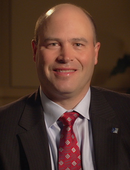

Geoffrey S. Baer, MD, PhD
OREF/National Stem Cell Foundation (NSCF) Research Grant in Stem Cells and Regenerative Medicine
Research Topic
Investigated whether mesenchymal stem cells could be genetically modified to produce factors that heal tendons by regeneration
Patient Impact
A new option for treating tendon injury that restores them to their original composition and mechanical properties without introducing scar tissue or increasing the risk of post-traumatic osteoarthritis
Regenerative Healing: Stem Cells for Service and Delivery
OREF grant recipient investigates regenerative options for tendon injuries
Jay D. Lenn, OREF contributing writer
Tendon and ligament injuries are common, and they can present both short-term morbidity and long-term changes in function. The healing process after surgery often does not restore the tendon or ligament to its original composition or mechanical properties. Rates of subsequent injury are high, and the risk of post-traumatic osteoarthritis is increased.
Several biological interventions, such as introducing growth factors at the site of surgical repair, have shown some benefits, but these treatments fall short of restoring tissues to their native state. Geoffrey S. Baer, MD, PhD, associate professor of orthopaedics and rehabilitation at the University of Wisconsin School of Medicine and Public Health in Madison, has been investigating a novel strategy to improve outcomes with biological factors.
Dr. Baer explained, "We're using mesenchymal stem/stromal cells that have been stimulated to increase the production of cytokines and receptor antagonists to enhance healing. We're trying to turn healing toward a more regenerative line, rather than heading down a line that results in scar tissue formation."
His research was funded by a 2015 Orthopaedic Research and Education Foundation (OREF)/National Stem Cell Foundation (NSCF) Research Grant in Stem Cells and Regenerative Medicine. This two-year grant provided $150,000 for innovative, translational adult stem cell research.
Promoting the good, inhibiting the bad
Dr. Baer's research is a convergence of multiple inquiries to promote regenerative healing of tendons and ligaments, combining promising treatments with a delivery vehicle that may optimize their benefit. A central character in these inquiries is the macrophage and its function in tissue repair.
Macrophages are the cleanup crew of the immune system, but different macrophage “phenotypes” play different roles. Macrophage polarization—the process that results in different macrophage phenotypes—guides the direction of tendon and ligament healing. Although a spectrum of macrophage phenotypes exists, they can be broadly categorized into the M1 and M2 phenotypes. Polarization to an M1 phenotype results in inflammation and scar formation while polarization to an M2 phenotype resolves inflammation and promotes tissue repair. Therefore, proteins that regulate macrophage polarization offer promising treatment strategies.
Two proteins that regulate macrophage phenotype include interleukin-4 (IL-4) and interleukin-1 receptor antagonist (IL-1Ra). Exposure of macrophages with IL-4 induces an anti-inflammatory M2 response. Conversely, exposure to IL-1Ra blocks the upregulation of the inflammatory cytokine, interleukin-1 (IL-1) and inhibits M1 macrophage polarization. Administration of either IL-4 or IL-1Ra could thereby potentially promote regenerative healing.
Dr. Baer’s group, including Drs. Connie S. Chamberlain and Ray Vanderby have performed a number of studies examining the effects of macrophage polarization on ligament and tendon healing using rodent models.1-7 When Dr. Baer's team treated the injury with injections of IL-4 or IL-1Ra at the time of surgical repair, the researchers observed less of an inflammatory response and an earlier healing response.2-4, 7 Dr. Baer noted, "Unfortunately, the effects were not long-lasting, possibly due to the short half-life of the proteins and their inability to remain localized at the treatment site." In the OREF/NSCF-funded study, he expanded this investigation with a novel delivery vehicle that might address these limitations: mesenchymal stem/stromal cells (MSCs).
Modifying stem cells for cargo
In the developing embryo, MSCs give rise to connective tissues throughout the body. In an adult, a small quantity of MSCs—found in bone marrow, fat and muscles—helps support tissue repair. Studies have shown that MSC therapy may improve healing by providing a direct contribution to tissue repair or by secreting proteins that promote regeneration.
Dr. Baer's group explored whether MSCs could not only provide their own therapeutic value but also enable a sustained, elevated production of IL-4 or IL-1Ra. Dr. Baer stated, "Our thought was that genetically modifying stem cells to overproduce or have a longer production of pro-healing factors may continue to push that line to enhance healing. The goal is to promote sustained healing and reduce scar formation.
Testing in mouse models of tendon injury
Dr. Baer and his colleagues tested the genetically modified stem cells in a mouse model of Achilles tendon rupture. Each mouse received the same surgically created injury, the injury was repaired with sutures, MSCs suspended in a collagen gel were injected at the injury site, the wound was sutured, and the limb splinted. Due to the injury models developing abscesses on the injection sites for IL-4, the researchers focused only on the effects of IL-1Ra. The study group for IL-1Ra treatment was divided into four subsets of mice receiving one of the following collagen gel injections:
- MSCs
- MSCs that overexpress IL-1Ra
- MSCs with IL-1Ra deleted
- No MSCs (control group)
The MSCs also were genetically modified to produce tomato red, a kind of built-in tracking device for monitoring the stem cells in vivo. The mice in each subset were euthanized at seven or 21 days after injury. The researchers then examined the healed tendons for their mechanical behavior, the composition and organization of the extracellular matrix, and macrophage polarization. Dr. Baer and his team analyzed the data. Their preliminary findings have indicated that deletion of IL-1Ra from the MSCs (i.e. loss in controlling inflammatory IL-1) resulted in tendons that were more inflammatory in nature. Conversely, delivery of MSCs overexpressing IL-1Ra to the injured tendon reduced inflammation for a longer period of time than other treatments tested. Further testing is currently underway to determine the mechanism of healing by these treatments.
More recently, Dr. Baer has expanded his research team at the University of Wisconsin-Madison to also include Drs. Peiman Hematti, John Kink, and Matt Halanski.6 Together, the team has examined the effects of macrophage delivery directly to the tendon injury. Here the team delivered a novel M2-like macrophage to the injured tendon and demonstrated significantly improved tendon healing as demonstrated via improved strength and less inflammation.
Supporting basic science and translational research
Dr. Baer noted the value of this investigation on more than one front: "We are exploring the potential of stem cell treatments to induce regenerative outcomes, elucidating the key aspects of ligament and tendon healing, and providing a methodology for future inquiry."
He added, "The OREF grant allowed us to take the research we did on a much smaller scale—just using injections of biological factors—and expand that into stem cells, where we see a lot of potential for healing. Without OREF, I think the ability to pursue these research interests would be extremely limited. We are really able to push toward translational research and to push toward patient care with this investigation."
References
1. Chamberlain CS, Leiferman EM, Frisch KE, Wang SJ, Yang XP, van Rooijen N, Baer GS, Brickson SL,Vanderby R. The influence of macrophage depletion on ligament healing. Connect Tissue Res 2011; 52:203-211.
2. Chamberlain CS, Leiferman EM, Frisch KE, Wang SJ, Yang XP, Brickson SL,Vanderby R. The influence of interleukin-4 on ligament healing. Wound Repair and Regeneration 2011; 19:426-435.
3. Chamberlain CS, Leiferman EM, Frisch KE, Duenwald-Kuehl SE, Brickson SL, Murphy WL, Baer GS,Vanderby R. Interleukin-1 receptor antagonist modulates inflammation and scarring after ligament injury. Connect Tissue Res 2014; 55:177-186. .
4. Chamberlain CS, Leiferman EM, Frisch KE, Brickson SL, Murphy WL, Baer GS,Vanderby R. Interleukin Expression after Injury and the Effects of Interleukin-1 Receptor Antagonist. PLoS One 2013; 8.
5. Chamberlain CS, Duenwald-Kuehl SE, Okotie G, Brounts SH, Baer GS,Vanderby R. Temporal Healing in Rat Achilles Tendon: Ultrasound Correlations. Ann Biomed Eng 2013; 41:477-487.
6. Chamberlain CS, Clements AEB, Kink JA, Choi U, Baer GS, Halanski MA, Hematti P,Vanderby R. Extracellular Vesicle-Educated Macrophages Promote Early Achilles Tendon Healing. Stem Cells 2019.
7. Chamberlain CS, Brounts SH, Sterken DG, Rolnick KI, Baer GS,Vanderby R. Gene profiling of the rat medial collateral ligament during early healing using microarray analysis. J Appl Physiol 2011; 111:552-565
